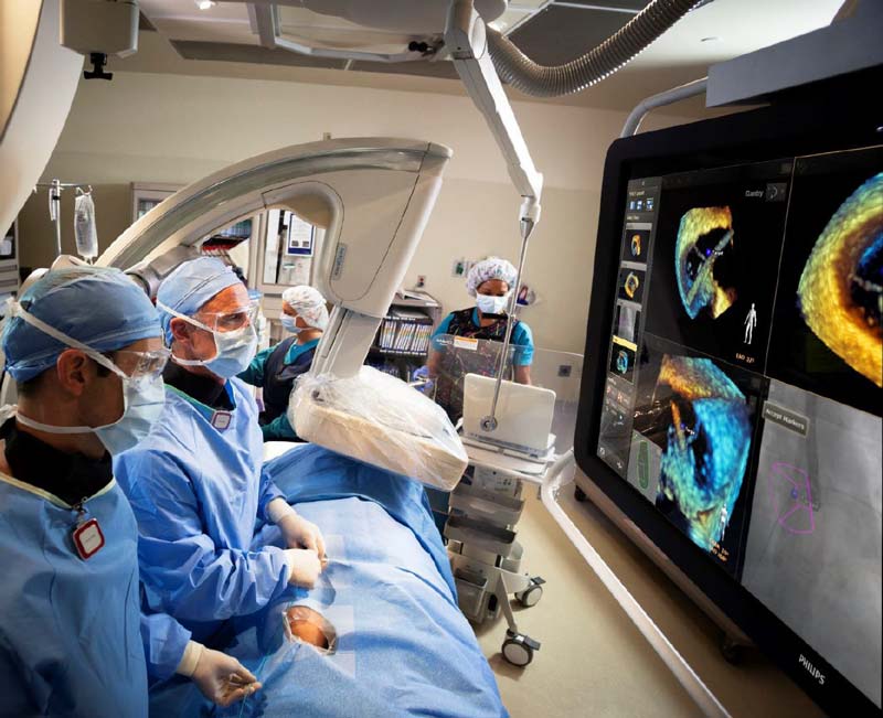
In this medical imaging use case, Philips Healthcare (PMS) will work on the analysis of X-ray (2D) and Echo (3D) image sequences that are acquired during the minimally invasive treatment of patients with structural heart disease (SHD). The X-ray images do not show the soft anatomy (the heart) of the patient but visualizes the in-body treatment device very well. The Echo images show the soft anatomy (the heart) nicely, but visualization of the treatment device is poor. The real-time analysis of the X-ray and Echo images enables the detection and tracking of the precise position and orientation or the treatment device and enables enhanced visualization of the device in the X-ray images and in the Echo images.
Automatic real-time image-based analysis of the X-ray and Echo data will support the doctor during the treatment, by providing an intuitive visualization of the device, which will simplify the clinical procedure, lead to shorter procedure times and allows for less skilled doctors to perform the same procedure.
PMS, in collaboration with imec-NL, is interested in the detection of a medical device called MitraClip, used in the treatment of a specific form of structural heart disease known as “mitral regurgitation” (MR). This is a leakage of mitral valve in the human heart. The objective of this use case is to be able to automatically detect a MitraClip in the X-Ray images alone by using a neural network technique known as YOLO.

